 Introduction
Dermatologic surgery is often performed in an outpatient surgical suite with local anesthesia, and moderate to advanced dermatologic procedures may be done without monitoring vital signs. A sound knowledge of the common dermatologic surgery complications is essential to a safe surgical practice. For example, identifying at-risk patients is key to preventing avoidable complications. Complications are to be expected in high volume practices. However, complications should be approached with well-rounded knowledge and managed accordingly. This chapter will present several of the most common surgical considerations for safe surgery and prevention and management of emergencies in dermatologic surgery.
The Golden Rule: Prevention
The main approach in reducing complications is prevention. Prevention of complications requires an excellent informed consent which consists of:
1. A clear definition of the procedure
2. A discussion of the key alternative therapeutic options
3. Education regarding the normal recovery time for different procedures
4. Education regarding the common and rare, major and minor side effects
Before any procedure in dermatologic surgery is performed, knowing how to treat the possible side effects is essential. An experienced dermatologic surgeon will take a moment to imagine the procedure from start to finish, including management of any and all potential side effects before actually starting the procedure. In this way, many side effects can be prevented. For example, if the patient is known to be on anticoagulation therapy and the possibility of a hematoma is anticipated, the intra-operative management of coagulation will be more conservative.
Finally, management of unforeseeable complications in dermatologic surgery also requires sensitivity regarding patients expectations and emotions. For example, bruising, which is an accepted side effect in many procedures, is stressful for patients. Dermatologic surgeons should be readily available for follow-up appointments if only to offer reassurance while minor side effects and complications are resolving. When major side effects occur, the ability to discuss the potential cause and the management plan with the patient, while accepting appropriate responsibility for that complication, is essential for successful management of dermatologic surgical complications.
Four of the most common complications in dermatologic surgery include hemorrhage, infection, dehiscence, and necrosis of the skin. Each of these will be discussed below.
Hemorrhage
Even the most skilled surgeon will encounter bleeding at some point in his or her practice. To minimize the clinical sequelae of excessive bleeding, the astute surgeon anticipates the potential difficulties and recognizes and manages problems as they arise. However, much of the battle is to be won off of the operating table with a thorough preoperative assessment, familiarization with current guidelines for withholding blood thinners, knowledge of management of delayed bleeding, and excellent pressure bandaging for the post-operative period of time.
Beyond excessive bleeding intra-operatively, postoperative bleeding can be both distressing for the patient as well have the potential for hematoma formation. A hematoma is a localized collection of blood outside of the blood vessel, usually in liquid form in the tissue. It appears as a deep purple, grape jelly-like nodule in or adjacent to the area of surgery. One common area for a hematoma is the infraorbital space occurring after surgery near or trauma to the infraorbital vessels. Another is the leg where superficial vessels may bleed or clot. A hematoma differs from an ecchymosis, or a bruise, which is due to the spread of blood under the skin—usually throughout the fat—in a thin layer. An ecchymosis may extend beyond the area of a hematoma, as in dependent areas or areas inferior to a surgical site, such as a wound on a cheek with an ecchymosis that extends around the jaw and submentally, or a wound on the forehead with periorbital bruising. Ecchymoses resolve after five to 14 days depending on the location. In contrast, a hematoma resolves much more slowly and often confers an increased risk of infection. The most common factor leading to hematomas in patients presenting for dermatologic surgery is concurrent anticoagulation therapy. Of the many types of anticoagulants, it is often aspirin, ibuprofen, or clopidogrel that contribute to increased risk of hemorrhage more than warfarin.
Assessing potential bleeding risk includes a thorough history of significant bleeding during prior low-risk surgical or dental procedures, common medical problems such as hypertension, alcoholism, and anxiety, and known medical conditions that contribute to abnormal coagulation, such as liver disease, renal dysfunction, and malignancies. Finally, it is imperative to obtain a medication history that includes medications the patient takes both daily and as needed, as well as the last date the patient may have taken each of these medications. Several common over the counter medications may have anticoagulation effects, including Alka-Seltzer, which contains aspirin, and Advil Cold and Sinus, which contains ibuprofen. Gingko biloba, garlic, ginseng, ginger, feverfew, vitamin E and saw palmetto have all been implicated in increased operative and post-operative bleeding. Many studies suggest waiting three days after the last dose of aspirin to reduce the risk of bleeding. However, the life span of a platelet is twelve days, the half life is six days, and one baby aspirin (81mg) effects all of the platelets in circulation. Therefore, one week is the ideal time to request patients avoid non-medically necessary supplements or anticoagulants. Excessive bleeding secondary to acquired abnormalities in coagulation or platelet function from medications or ingested substances is surprisingly common. (TABLE 14.1) For example, alcohol decreases vasoconstriction and impairs coagulation of platelets. Furthermore, various dietary supplements and alternative medicines have the potential to alter the coagulation cascade and should therefore also be taken in to account when obtaining a medication history.
The decision to discontinue non-steroidal anti-inflammatory medicines (NSAIDs) is important. (TABLE 14.2) Many take NSAIDs for non-medical but necessary reasons such as comfort. However, a significant proportion take NSAIDs or other anticoagulant/antiplatelet medications for medically necessary reasons such as prevention of cardiac and/or cerebrovascular events, making the decision to discontinue such medications moot. Studies comparing hemorrhagic complications have generally not shown an increased risk of severe hemorrhagic complications in patients on anticoagulants versus those who are not.,,, This topic has been debated in the dermatologic surgery literature, specifically regarding warfarin, aspirin, and NSAID use, and there is a general consensus that continuation of these anticoagulants is associated with a very low risk of complications and is not statistically increased compared with patients in whom the same medications are discontinued.,,
Despite the current literature, the decision about whether to discontinue anticoagulation remains, and one must consider the real and increased risk of bleeding and hemorrhage with the lower but potentially life-threatening risk of a thrombotic event should an anticoagulant be temporarily discontinued.,,, For instance, the risk of a thrombotic event from discontinuing anti-thrombotic medications may outweigh the risks of intra-operative bleeding. Kirkorian et al surveyed current practice regarding perioperative management of anticoagulant therapy amongst dermatologic surgeons finding 87% discontinue prophylactic aspirin therapy, 37% discontinue medically necessary aspirin, 44% discontinue warfarin, 77% discontinue NSAIDs, and 77% discontinue vitamin E therapy perioperatively at least some of the time. Although clopidogrel was not surveyed in this article, 78 physicians included comments about the management of this agent. Bordeaux et al concluded that warfarin or clopidogrel increased bleeding risk, but these medications should be continued to avoid adverse thrombotic events.,
Should there be excessive bleeding during surgery, the use of proper bandages after surgery will help prevent hematoma formation. In our practice we use condensed cotton dental roles under 4×4 gauze followed by Hypafix™ paper tape which has an elastic property to create a pressure dressing that remains on the site for 24 hours following Mohs or excisional surgery. In addition, we recommend that those on anticoagulant medications or who present with excessive bleeding during surgery employ ice packs for 20 minutes every hour for the first six hours. Patients are given a detailed, written handout with instructions on how to manage excessive bleeding including application of ice and pressure for 20 minutes continuously without removing the pressure to examine the area. Patients are advised to call or return to the office promptly if they have concerns such as a warm, expanding and possibly painful mass, or especially if involving the periorbital or cervical spine areas.
The primary method to treat an expanding hematoma is 1) partial or complete opening of the surgical wound, 2) identifying the culprit vessels and stopping bleeding by suture ligation or electrosurgery, 3) and either a full-layer re-closure of the wound or allowing the wound to heal by secondary intention. The latter is considered if there is a high risk of more bleeding or the wound is contaminated.
Clinical Pearl: To prevent a hematoma intra-operatively, take time to implement proper positioning of the patient, the surgeon, and the instrument tray. Something as simple as proper positioning may yield a better field for the dermatologic surgeon to visualize the source of bleeding, and ensure effective perioperative hemostasis.
Infection
Surgical site infections (SSIs) after dermatologic surgery cause pain, prolong healing, may result in altered cosmesis, and lead to excessive use of antibiotics. Signs and symptoms of wound infection usually present 4-6 days post-operatively with erythema, pain, warmth and swelling at the wound site. If cellulitis and fever are present, prompt institution of oral antibiotic therapy and close follow-up care are adequate. However, if the wound is fluctuant, if there is purulent exudate or inflammatory edema, or if systemic symptoms are present, the wound must be opened, lavaged with sterile sodium chloride solution and packed with iodoform gauze. Cultures and gram stains should be performed, and broad-spectrum antibiotics should be instituted while awaiting results. Patients should be closely monitored to evaluate clinical response.
Despite the potential for SSI, antimicrobial prophylaxis is rarely appropriate for dermatologic surgery. Reported infection rates for Mohs micrographic surgery (MMS) range from less than 0.7% to 3.5% and occur most frequently on the legs., Dermatologic procedures seldom cause bacteremia, and they have been implicated in only an extremely small fraction of cases of endocarditis or infections of vascular grafts or orthopedic prostheses. According to the newest guidelines, systemic prophylactic antibiotics are rarely indicated in patients undergoing dermatologic surgery, even those who would otherwise require prophylaxis for dental procedures. (TABLE 14.3) Because wound infections following dermatologic surgery are uncommon, usually mild, and generally easily treatable, systemic antimicrobial prophylaxis is not indicated to prevent postoperative wound infections either. Topical antibiotic ointments for that purpose are also ineffective, yet often used regardless.
Although the overall trend in the literature supports decreased use of antimicrobials in dermatologic surgery as a whole, it is important to know which situations still warrant antibiotics. Patients at high risk for wound infection should receive prophylactic antibiotics chosen to cover the organism most likely to cause infection. Staphylococcus aureus is the most common agent of skin wound infections. However, Streptococcus viridans is commonly found in oral flora, and Escherichia coli is present near the gastrointestinal and genitourinary tracts. For legs with granulating wounds, common causes of postoperative infections are from E. coli from above. For this reason, this author instructs all patients to use Hibiclens surgical scrub from the groin down. Pseudomonas species are a common pathogen of the external ear. If the patient has chondritis of the ears post-operatively, it may not be a true infection. However, patients are often put on ciprofloxin 500mg by mouth twice a day for seven days to cover for Pseudomonas. Additionally, any time a patient has a laser procedure, photodynamic therapy or surgical procedure around the lips, it is important to get a full history of herpes infections—ask about fever blisters- and institute prophylaxis with acyclovir or a related medication. This author uses Famvir 500 mg by mouth twice daily for 3 days for anyone with a history of HSV, or for anyone receiving ablative procedures such as CO2 laser resurfacing.
As mentioned in Table 2, dermatologic surgery is not considered in the AHA guidelines for endocarditis prophylaxis. In one review, the authors did not recommend routine prophylaxis for high-risk patients undergoing procedures of less than 20 minutes duration on intact skin. (TABLE 14.3) Patients at high risk for endocarditis should receive prophylaxis in cases where there is infected skin, breach of oral mucosa, or non-infected site at high risk for surgical site infection,. This prophylaxis should be administered 30-60 minutes prior to performing the procedure to allow the antibiotic to be present in the blood at the time of the initial incision. Appropriate specialists should be consulted—and documented-for each procedure performed on patients with orthopedic prostheses, those with ventriculoatrial and peritoneal shunts, and in any case where the surgeon is not comfortable making the decision alone. A 2008 advisory statement on antibiotic prophylaxis is the most recent and widely used reference algorithm for dermatologic surgeons.
Clinical Pearl: Three main factors are essential in the decision making process regarding antibiotic prophylaxis.
Infection control measures including active surveillance of SSI, implementation of a protocol for dealing with SSI, compliance observations and instruction/training of healthcare workers, clipping surrounding hair instead of shaving, and following the often-times confusing perioperative antibiotic prophylaxis guidelines are essential measuresin the prevention of SSI. Though preoperative MRSA screening and implementation of a decontamination protocol appears to decrease postoperative MRSA wound infections after Mohs surgery, the clinical efficacy and cost effectiveness of this screening has yet to be determined. Finally, infection control includes safety of the surgical team from exposure to HIV or Hepatitis C virus. Vigilant infection control for both the patient and the team is critical to surgical safety.
Necrosis
Necrosis of tissue occurs secondary to ischemia. Any condition that results in a decrease of oxygenated blood flow to the wound has the potential to cause necrosis. Too much tension on wound edges, excessive suturing and undermining, and superficial undermining all may result in tissue compromise and decreased blood flow to the distal wound margins. Hematomas may create excessive tension on the wound, increasing the risk of necrosis. Flaps and grafts with a length-to-base ratio of greater than 4:1 are at high risk of necrosis because of the tenuous circulation at the wound edges. Location of the wound is also an important consideration; high-risk areas include the lips, ears, nose and skin grafts. Additionally, anesthesia type, amount and addition of epinephrine all affect circulation around the wound and can contribute to ischemia.
Cigarette smoking results in vasoconstriction, increased blood viscosity, hypoxia, increased platelet aggregation, and clots. Smoking more than 1 pack of cigarettes per day increases the risk of necrosis by 3-fold compared to never smokers, ex-smokers, and smokers of less than 1 pack per day. When necrosis does occur, the area involved tends to be greater in smokers compared to those who have never smoked. Stopping or decreasing smoking for at least 2 days prior to surgery and for at least 1 week post-operatively can potentially reverse some of the negative effects of smoking on the microvasculature.
When necrosis occurs, treatment is conservative. Except in cases of hematoma formation and infection, the full extent of the necrosis should be allowed to manifest. Debriding the wound prior to this may injure viable tissue. When the eschar is freely separable from the wound bed, careful and sharp debridement may be performed, and the wound may be allowed to granulate. Necrotic wounds are at higher risk for infection and the surgeon should observe the affected area carefully and frequently, and consider systemic antibiotics.
Dehiscence
Wound dehiscence is a surgical complication in which the layers of a wound unintentionally re-open. Wound dehiscence may be caused by anything that causes excessive tension on the wound edges. This includes inadequate undermining or putting undo strain on the wound after surgery. When wounds are located in highly mobile or high tension areas such as the back, shoulders or legs, the risk of wound dehiscence is higher. Underlying conditions such as Ehlers-Danlos syndrome, allergic contact dermatitis to adhesive or topical antibiotics, infection, spitting sutures, or obesity can also increase risk for wound dehiscence. Other risk factors include smoking, previous scarring or radiation, cancer, recent use of Accutane, or chronic use of corticosteroids.
Dehiscence can be prevented by adequately undermining the tissue and using as much tissue reservoir as necessary to close the wound without tension. The patient should be instructed to avoid heavy lifting or extra activity, maintain adequate nutrition to speed wound healing, control underlying conditions that might alter wound healing (such as uncontrolled diabetes), and avoid prednisone use during the healing period. Because hematomas may also contribute to wound dehiscence, appropriate post-operative bandaging is important, as is perioperative bleeding management.
If wound dehiscence occurs, the surgeon has several options, including allowing the wound to granulate on its own or re-cutting and re-suturing the wound edges. For wound dehiscence on the face, there are several options based on the nature, time, and condition of the wound after dehiscence. For instance, if dehiscence is caused by trauma (e.g. a patient trips and falls and hits a graft site on the nose) or premature suture removal (e.g. the wound needs more than the standard 7 day period to heal), refreshing the wound edges and re-suturing is acceptable if there is no evidence of infection or the edges look intact. If signs of infection are present, appropriate antibiotic therapy is recommended. Areas such as the temple are known to heal poorly due to poor circulation. Allowing small dehiscent areas to granulate for 2-6 weeks is often the best course of action. The skin will heal, there will be less serous fluid, and the wound edges will be more amenable to refreshing and re-suturing. Too much intervention, or premature intervention, may worsen the cosmetic outcome. However, deeper dehiscence involving muscle, such as wedge reconstructions of the lip may require immediate intervention. One tip: if Vicryl™ sutures were rejected prematurely resulting in dehiscence, switch to Monocryl™ when reconstructing the wound. Because Monocryl is monofilament and less inflammatory, it has a longer duration and lower probability of spitting suture. In summary, for most cases of wound dehiscence, appropriately manage infection, excess moisture, and allow granulation to occur unless the dehiscence involves deeper layers where cosmesis will be difficult to achieve with a delayed intervention.
Other Dermatologic Procedural Emergencies:
1. Laser Emergencies
Today in dermatology, hundreds of different laser and light devices exist for various indications, including excessive hair, superficial and deep vascular lesions, tattoos, lentigines, birthmarks, cutaneous aging and laxity, and psoriasis. In many cases, laser complications result from collateral damage created when energy intended for the target chromophore is nonselectively diffused and absorbed by surrounding tissues. The theory of selective photothermolysis aims to reduce laser complications by selectively heating a target chromophore while sparing the non-intended but nearby targets. For example, melanin selectively absorbs photons in the 532nm wavelength creating heat, resulting in pigment destruction, while sparing the surrounding cells. Mild, short-term complications of laser treatment include erythema, edema, urticaria, crusting or erosion, ecchymoses, blistering and infection, such as reactivation of herpes simplex virus (HSV). More moderate, longer-lasting complications include pigmentary changes, visible lines of demarcation, burns, textural abnormalities, or delayed reepithelialization. Severe, long-term complications include permanent dyspigmentation, scarring, and ocular damage. The complications noted above are devastating, but are not emergent. Fortunately, there are few, emergencies from laser treatment other than ocular damage. All potential side effects of a particular device should be discussed and provided in writing ahead of time to a patient, along with instructions for appropriate cutaneous care following laser treatment. Additionally, if the patient has a history of HSV, prophylaxis should be instituted prior to the procedure. Close follow up is essential in the management of these potentially upsetting, but non-emergent problems.
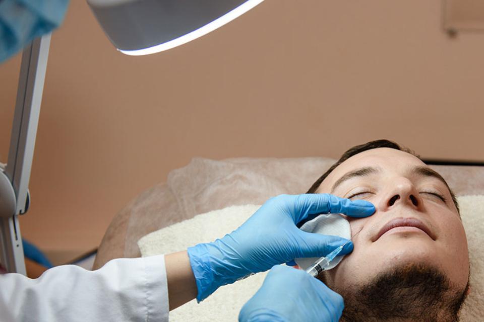
2. Filler Emergencies
Inappropriate placement of filler such as superficial placement is one of the most frequent reasons for patient dissatisfaction. Some fillers, including hyaluronic acid, are reversible with hyaluronidase, and these complications can be easily corrected. Hypersensitivity to current FDA approved products is rare and can most often be managed with anti-inflammatory agents. Infection is also uncommon and is managed with antibiotics or antivirals, depending on the etiology. The most concerning complication is necrosis.
The risk of skin necrosis following injection of filler is increased in certain potential danger zones, including the glabella, nasal skin, temple, alar groove and even the lip., The most common of area for filler-related necrosis is the glabella. Major facial vessels of concern during soft tissue augmentation are the superior and inferior labial arteries, both branching off the facial artery at the angle of the mouth; the angular artery, the terminal branch of the facial artery; the lateral nasal artery, branch of the angular artery; the dorsal nasal artery, which forms an anastomosis with the angular artery; the supratrochlear artery, one of the terminal branches of the ophthalmic artery; and the superficial temporal artery, arising from the external carotid artery.
Several treatment algorithms for impending facial necrosis due to filler injection have been proposed in the literature.,,,, If tissue blanching is observed, injection should be stopped immediately. Warm gauze (to facilitate vasodilation) and tapping (to potentially break up product) are then recommended. If the filler was an HA, immediate injection of hyaluronidase should be used. The authors recommend injection of a 1:1 volume of hyaluronidase to HA in the zone of ischemia or necroses. Typically, a range of 8u-16u vitrase 0.5mL of HA will dissolve the cosmetically imperfect swelling. For emergency situations when occlusion or ischemia is suspected, use more. No absolute maximum dose has been published but this author has used 50u hyaluronidase in the area of the alar crease to correct a 0.2mL compression induced ischemia of the ipsilateral ala. Per the literature, it is exceedingly rare to dissolve native HA with hyaluronidase. Immediate use of nitroglycerin paste to facilitate vascular dilatation and blood flow to the area should be applied. Additionally, anticoagulants such as aspirin and non-steroidal anti-inflammatory agents, as well as vitamins that inhibit platelet aggregation, should be encouraged in any protocol to treat cutaneous tissue ischemia.
Given that hyperbaric oxygen has proven successfulin healing of nasal tip grafting in cases of cancer or trauma reconstruction, this author suggests administration of oxygen via nasal cannula, face-mask or in the form of hyperbaric oxygen for ischemia secondary to cosmetic filler injection., For cases with severe, unremitting swelling, oral antihistamines may also be employed. Finally, treatment with low molecular weight heparin (5000 IE daily) has been reported to treat impending necrosis after hyaluronic acid injection in the treatment of frown lines of the glabella due to occlusion or compression of the supratrochlear vessels..
3. Botox Emergencies
There are many neurotoxins approved by the FDA in use today. Botox acts via prevention of the release of acetylcholine at the presynaptic nerve terminal, thereby stopping the electrical impulse and blocking muscular movement. Complications of botox include:
• Overcorrection
• Undercorrection
• Asymmetric result
• Upper eyelid ptosis
• Dysphagia
• Neck weakness
• Perioral droop
• Bruising
• Headache
• Intravascular injection
• Globe perforation
• Diplopia
Particularly distressing can be true eyelid ptosis, which occurs when botox is injected or diffuses into the levator palpebrae superiorus muscle. Though temporary, this effect can be distressing for the patient and can impact their quality of life. To avoid this complication, all injections should be at least 1 cm above the bony orbital rim. If this complication occurs, Iopidine (apraclonidine) 0.5% drops may be used to stimulate Muller’s muscle, providingsome lid opening to partially alleviate the ptosis.
Similar to those complications encountered with laser treatment, the adverse effects of Botox can be distressing to the patient, but few, if any, represent a true emergency.
4. Chemical Peel Emergencies
Common chemical peel side effects include stinging, redness, and desquamation of the skin. These side effects should be expected by any patient who undergoes a chemical peel, as the mechanism of the peel is controlled skin injury in an attempt to remodel the epidermis., Chemical peels can target the superficial, medium or deep layers of the epidermis, depending on the type of chemical used and the desired outcome. Other complications of peels include contact dermatitis, milia formation, infection, acne or rosacea flare, and pigmentary changes. It is important to discuss the common side effects prior to performing the procedure on any patient. Additionally, certain medications may potentiate the effects of the peel and are thus necessary to be aware of prior to performing the procedure.
The most superficial chemical peels include alpha-hydroxy acids (AHA), such as glycolic acid, and these peels are associated with minimal side effects. The two major side effects of AHA peels are irritation and sun sensitivity, which are transient. Medium depth peels include trichloroacetic acids (TCA). Side effects are similar to AHA peels but last longer. The deepest peels include phenols, and can be painful requiring local anesthetic or sedation. Healing time is longer- usually two to three weeks- and the most consistent side effect is pigmentary alteration (hypo- or hyperpigmentation). Other serious side effects of phenol include scarring, infection, as well as rare cardiac arrhythmias and temporary laryngeal edema. Patients who have impaired liver or kidney function are most at risk of phenol toxicity, as phenol is absorbed through the skin and subsequently metabolized by the liver and excreted by the kidneys. In addition, the size of the treatment areas, rather than the concentration of the phenol, determines the extent of cutaneous absorption. To minimize these serious complications, cardiac monitoring during the procedure and in the immediate recovery period, as well as pausing treatment for 15 minutes between each cosmetic unit is recommended. Regarding laryngeal edema, heavy smokers seem to be most at risk for this rare but serious complication.
One very real emergency of any chemical peel is a burn of the eye from an accidental splash. Cover the eyes with proper protection such as tape or stickers. Arrange the procedure tray to avoid accidental spillage; always label of the peel container to ensure the proper product, and place the original bottle on the counter as opposed to the Mayo stand, where it can more easily spill. Additionally, a clearly marked eye wash station is essential should a splash occur. Furthermore, immediate evaluation by an ophthalmologist or transfer to the emergency department is warranted should an accident occur.
Clinical Pearl: To avoid complications of chemical peels:
Conclusion
Dermatologic surgery is an art that involves complex procedures involving excisions, repairs, injections and devices. Thorough understanding of the anatomy and pathophysiology of the skin, proper patient evaluation, appropriate selection of procedures based on each patient, and divulging potential complications of each procedure are essential to achieving optimal results and minimizing complications in cutaneous procedures.