 To better understand the skin and its behavior we must first look at its component parts.
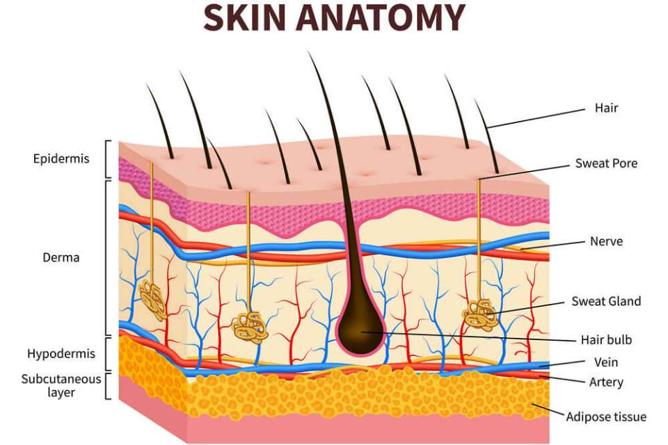
The skin is composed of 3 layers: the epidermis is the outermost layer and is composed of keratinocytes or skin cells that form the “bricks” of our skin’s barrier. The functions of the epidermis are protection from environmental insults (like ultraviolet light and toxins), prevention of dryness, and immune surveillance. The base of the epidermis is called the basal layer – it contains the cells that replicate in order to replace the epidermis every month. Mixed in between keratinocytes of the epidermis are pigment cells, called melanocytes, that give skin its characteristic color. These cells become activated with ultraviolet exposure found in sunlight. The result of this activation is two-fold – 1) melanocytes produce more melanosomes, envelopes that contain brown melanin pigment, and 2) the increased transfer of these melanosomes to adjacent skin cells. The result is freckling or sun-spots that can significantly impact appearance.
Beneath the epidermis is the dermis, composed mostly of collagen but also adjunctive structures like hair follicles and sweat glands. Sebaceous glands are found next to hair follicles and produce sebum, a combination of natural lipids that coat the skin’s surface and provide a protective nourishing role. Sweat glands function to help regulate temperature through evaporation and cooling. Their ducts pass through the dermis and epidermis to empty directly onto the skin’s surface. The dermis also contains vital blood vessels and nerves which traverse the collagen network there. Within the dermis also lies a protein, elastin, that provides cutaneous elasticity and fibroblasts, the cells that produce more collagen. The function of the dermis is temperature regulation though the secretion of sweat to the skin’s surface and the regulation of blood flow to the area. The dermis also provides mechanical protection for the adenexal structures discussed above.
Below the dermis, lies the subcutis which holds fat and larger blood vessels. Fat is arranged into lobules that are several millimeters wide. These lobules are divided by thin wisps of tissue that contain blood vessels and nerves. The subcutis acts as a heat insulator and also provides protection from mechanical trauma.
Most skin disorders begin with a malfunction of one of the anatomical components discussed above. For example, acne occurs when a hair follicle is occluded, leading to an over-abundance of bacteria behind that plug. Similarly, rosacea occurs when blood vessels that traverse the dermis become dilated and give the skin that red color so characteristic of this condition. A better understanding of your skin begins with a clear appreciation of its anatomy described above.