 Vasculitis is defined as inflammation of blood vessel walls. There are many types of vasculitis, lending to its variable clinical presentation, with or without systemic involvement. The diagnosis of specific vasculitides can be difficult, given much overlap in clinical manifestations, serum laboratory tests, and tissue histopathology. In addition, it is essential to differentiate benign, self-limited forms of vasculitis from those that may be life-threatening.
Classification
Several classification systems have been used. One common system is based on vessel size. Large vessels encompass the aorta, as well as its branching large arteries and veins that are directed toward major body regions, such as carotid arteries and its branches. Giant cell arteritis and Takayasu arteritis mainly involve these vessels. These large arteries are not found in the skin although skin manifestations can be seen in these vasculitides. Medium vessels are the main visceral (renal, hepatic, coronary, and mesenteric) vessels; these vessels are implicated in entities such as polyarteritis nodosa and Kawasaki disease. They may also be involved in Wegener’s granulomatosis, Churg-Strauss syndrome, microscopic polyangitis, and cryoglobulinemic vasculitis. Small vessels are arterioles, venules, and capillaries. These vessels are involved in many types of vasculitis such as cutaneous leukocytoclastic vasculitis, Wegener’s granulomatosis, Churg-Strauss syndrome, microscopic polyangitis, Henoch-Schonlein purpura, and others accounting for the majority of visible cutaneous disease.
Two classification schemes are those of the American College of Rheumatology (ACR) and the Chapel Hill Consensus Conference (CHCC). The ACR criteria of 1990 are a set of clinical and histological features that classify vasculitides. The CHCC definitions of 1992 provide histological definitions for 10 types of vasculitis. Both of these classification schemes were designed as research tools. CHCC definitions are based on histology and have limited value for clinical diagnosis. The ACR criteria are more amenable to clinical diagnosis, but these criteria have a positive predictive value of only 17-29% for the diagnosis of specific vasculitides. An expert group from the European League Against Rheumatism (EULAR) has recently suggested that new diagnostic and classification criteria should be developed with more consideration given to current diagnostic testing. While this group did not propose a new classification system, they proposed 17 points to consider in the development of such a system. These points included biopsy, laboratory testing, radiologic testing, nosology, definitions, and research agenda.
Pathogenesis
Vasculitis may be caused by a Type III hypersensitivity reaction which leads to deposition of immune complexes in blood vessel walls. This deposition is an early event in pathogenesis and is followed by activation of complement. Complement may directly damage the endothelium and also provides a chemotactic signal for the recruitment of inflammatory cells. The immune complexes themselves are also thought to activate polymorphonuclear leukocytes (PMNs) and increase production of tumor necrosis factor-alpha (TNF-alpha), Fas ligand, and perforin. Vessel damage may occur during diapedesis of PMNs from the luminal side of the vessel wall.
In the anti-neutrophil cytoplasmic antibody (ANCA)-associated vasculitides (Wegener’s granulomatosis, Churg-Strauss syndrome, and microscopic polyangitis), antibodies directed against myeloperoxidase (MPO) or proteinase 3 (PR3) are present. Anti-MPO antibodies increase leukocyte adhesion to endothelial cells and migration into renal and pulmonary tissues. These antibodies also activate MPO; this generates an oxidative stress which leads to damage of endothelial cells. PR3 bound to the neutrophil membrane plays a proinflammatory role, and anti-PR3 antibodies increase the expression of membrane-bound PR3 in neutrophils during cell adhesion. Anti-PR3 also activates epithelial cells and increases epithelial cell production of proinflammatory cytokines (interleukin (IL)-6, IL-8, monocyte chemoattractant protein 1, and TNF).
Cell-mediated immune responses may also play a role in giant cell arteritis, Takayasu’s arteritis, Wegener’s granulomatosis, and Churg-Strauss syndrome. CD4+ T cells are activated by antigens in vessels walls or in the circulation. These cells produce chemotactic cytokines which recruit monocytes. Monocytes mature into macrophages which produce lysosomal enzymes that damage endothelial cells.
The emergent nature of vasculitis
Vasculitis is an emergency because of systemic complications which can be life-threatening. Renal, gastrointestinal, pulmonary, and cardiac complications can be seen with specific vasculitides. Urgent diagnosis of vasculitis can ensure prompt initiation of appropriate treatment.
Types of vasculitis
Cutaneous small vessel vasculitis
Cutaneous small vessel vasculitis, also known as cutaneous leukocytoclastic angiitis, is caused by inflammation in post-capillary venules. Lesions often occur in crops on the lower extremities. Palpable purpura is the classic clinical finding, but macules, urticarial lesions, and vesicles can also be seen. Once mature, these lesions do not blanch with diascopy. Patients may develop fever, malaise, arthralgia, and myalgia. Underlying causes include infections, medications, foods (gluten, milk proteins), chemical exposure (petroleum products, insecticides), autoimmune disease, and malignancy. Despite the numerous causes, no specific etiologic factor is identified in up to 60% of patients.
Henoch-Schonlein purpura
Henoch-Schonlein purpura (HSP) is a small vessel vasculitis that is associated with immunoglobulin A immune complex deposition. It accounts for approximately 10% of cases of small vessel vasculitis. HSP is the most common type of vasculitis in children. It classically occurs in boys 4 to 8 years of age. Peak incidence is in winter months, and patients often have an upper respiratory tract infection 1 to 2 weeks prior to the development of symptoms. A recent study by Weiss et al has shown temporal association between hospitalizations for Group A beta-hemolytic streptococcus, Staphylococcus aureus, and Parainfluenza and hospitalization for HSP. Despite this temporal association, a causal role for these organisms has not yet been established.
The classic tetrad of HSP includes palpable purpura, arthritis, nephritis, and gastrointestinal tract involvement with abdominal pain or bleeding. All patients with HSP have skin involvement. In a study of 100 children with HSP, 83% had arthritis, 63% had abdominal pain, 33% had gastrointestinal bleeding, and 40% developed nephritis. Adults commonly present with joint or kidney involvement, but gastrointestinal manifestations are less common. Adults may be more refractory to treatment than children.
Palpable purpura is the classic cutaneous finding. Lesions may begin as macular erythema or urticaria that develops into nonblanching erythematous macules and papules. Lesions are symmetrically distributed, and dependent areas such as the lower extremities and buttocks are the most common sites of involvements. Individual lesions generally resolve within 10 to 14 days. While this condition is self-limited in most patients, the physician must take care to evaluate for renal involvement. Long term renal impairment is seen in 1.8% of all children with HSP and in 19.5% of children who develop nephritic or nephrotic syndrome. In a study by Coppo et al, end stage renal disease was seen in 15.8% of adults and 7% of children with HSP nephritis. The use of systemic therapy to decrease the likelihood of renal complications remains controversial. In one study by Ronkainen et al, prednisone did not prevent renal disease from occurring although it led to faster resolution of disease once it occurred. Prednisone was also effective in decreasing the intensity of abdominal pain and joint pain in this study. A more recent study from these authors compared the outcomes of patients 8 years after treatment with prednisone or placebo at disease onset. This study found no beneficial effect from early prednisone treatment, and the authors concluded that prednisone should not be routinely used.
Urticarial vasculitis
Urticarial vasculitis is a small vessel vasculitis that is seen in 1 to 10% of patients with chronic urticaria. It has a predilection for women and is most common in the fourth or fifth decades.
Patients with urticarial vasculitis have wheals and erythematous plaques that persist in the same location for more than 24 hours. Patients often complain of burning pain in these lesions. These features contrast with the evanescent pruritic lesions seen in classic, allergic urticaria, which is IgE-mediated. Lesions of urticarial vasculitis favor the proximal trunk and extremities. Petechiae or purpura are often seen, and lesions frequently resolve with postinflammatory pigmentary alteration.
Normocomplementemic urticarial vasculitis tends to be limited to the skin. It is often self-limited and may be regarded as a subset of cutaneous small vessel vasculitis. Low complement levels are seen in 18 to 32% of patients with urticarial vasculitis. Patients with hypocomplementemic urticarial vasculitis have more severe disease with involvement of the joints (50% of patients), the gastrointestinal tract (20% of patients), and the airways with asthma and obstructive airways disease (20% of patients). Some patients with severe disease may be considered to have hypocomplementemic urticarial vasculitis syndrome (HUVS). Patients with this syndrome may have renal involvement with glomerulonephritis or ophthalmologic involvement with uveitis or episcleritis in addition to the above symptoms. All patients with this syndrome have anti-C1q precipitins, and 24% may have anti-double stranded deoxyribonucleic acid (dsDNA) antibodies. There are overlapping features of HUVS and systemic lupus erythematosis (SLE). Fifty percent of patients with HUVS are subsequently diagnosed with SLE; HUVS is seen in 7 to 8% of patients with SLE. Another syndrome associated with urticarial vasculitis is Schnitzler syndrome. This syndrome is characterized by urticarial vasculitis, immunoglobulin M monoclonal gammopathy, fever, lymphadenopathy, arthralgia, bone pain, hepatosplenomegaly, elevated erythrocyte sedimentation rate, leukocytosis, and abnormal bone radiology studies. This syndrome also has overlapping features with SLE. Urticarial lesions in SLE may be more pruritic and difficult to control than those of Schnitzler syndrome. Schnitzler syndrome also commonly shows a neutrophilic leukocytosis while SLE may have neutropenia and positive antinuclear antibody (ANA). Schnitzler syndrome is benign in most patients although there is a risk of development of lymphoproliferative disorders.
Cryoglobulinemic vasculitis
Cryoglobulinemic vasculitis affects both small and medium-sized vessels. Cryoglobulins are immunoglobulin molecules that precipitate in cold temperatures. This precipitation occurs in vitro and may be due to intrinsic characteristics of the immunoglobulin components. Type I cryoglobulins are monoclonal immunoglobulin (Ig) M and less commonly IgG. These cryoglobulins are associated with hematologic conditions such as multiple myeloma or Waldenstrom’s macroglobulinemia. Type I cryoglobulins are associated with cold-induced vasculopathy but are not associated with vasculitis. Vasculopathy refers to vascular occlusion without blood vessel wall inflammation. The cryoglobulins associated with vasculitis, types II and III (the mixed cryoglobulins), are monoclonal IgM directed against polyclonal IgG and polyclonal IgM directed against polyclonal IgG, respectively. Fifteen percent of patients with cryoglobulins have cryoglobulinemic vasculitis. This vasculitis results from deposition of immune complexes in blood vessel walls with subsequent complement activation and inflammation.
In a study of 443 patients with cryoglobulinemia, underlying infection was seen in 75%, autoimmune disease in 24%, and hematologic disease in 7%. Hepatitis C virus is the most common infection associated with cryoglobulinemia. Hepatitis B and human immunodeficiency virus are less frequently associated. Associated autoimmune diseases include Sjögren’s syndrome, systemic sclerosis, SLE, rheumatoid arthritis (RA), and primary antiphospholipid syndrome. Hematologic associations include non-Hodgkin’s lymphoma, Hodgkin’s lymphoma, chronic lymphocytic leukemia, chronic myeloid leukemia, multiple myeloma, and myelodysplasia.
Cryoglobulinemic vasculitis often presents with palpable purpura localized to the lower extremities. Other cutaneous manifestations include erythematous papules, dermal nodules, ecchymoses, skin necrosis, bullae, urticarial lesions, ulcers, and livedo reticularis. In contrast to the vasculopathy associated with type I cryoglobulins, cold temperatures induce lesions in only 10 to 30% of cases. Common extracutaneous manifestations include joint involvement, peripheral neuropathy, and renal involvement. Patients may also have weakness, fever, lymphadenopathy, central nervous system, pulmonary, and gastrointestinal tract involvement. Central nervous system involvement may lead to devastating complications such as cerebral ischemia, spinal cord complications, or cranial nerve palsy.
Detection of cryoglobulins requires careful handling of specimens. Two red-topped tubes (without anticoagulant) must be drawn and transported to the laboratory at 37 degrees Celsius (C). This may be achieved by submerging the tubes in warm water or carrying the specimen in the axilla. The sample is allowed to clot at 37 degree C for 1 hour prior to centrifugation. Type I cryoglobulins may precipitate within 24 hours, but it may take several days for precipitation of mixed cryoglobulins. Many labs observe specimens for up to 7 days. Rheumatoid factor can serve as a surrogate marker for Type II and III cryoglobulins because this test looks for IgM directed against IgG in the blood.
Microscopic polyangitis
Microscopic polyangitis is a necrotizing vasculitis that affects small and medium-sized vessels. It is most frequently associated with perinuclear-antineutrophil cytoplasmic antibody (pANCA; anti-myeloperoxidase antibody) although cytoplasmic-antineutrophil cytoplasmic antibody (c-ANCA; anti-proteinase 3) is less frequently seen. p-ANCA has a sensitivity of 58% and specificity of 81% in the diagnosis of microscopic polyangitis; c-ANCA has a sensitivity of 23%. It may be slightly more common in men, and the average age of onset is 57 (37).
The first symptoms of microscopic polyangitis are typically fever, weight loss, arthralgia, and/or myalgia which may begin months to years before other disease manifestations. Palpable purpura is the most common cutaneous manifestation and is seen in 45% of patients at diagnosis. Other skin findings include splinter hemorrhages, nodules, livedo reticularis, and palmar erythema. Renal involvement, seen in 79-90% of patients, manifests as a focal segmental necrotizing glomerulonephritis. Pulmonary involvement occurs in 25 to 50% of patients, and pulmonary hemorrhage is seen in 12 to 29% of patients.
Microscopic polyangitis must be differentiated from other causes of pulmonary-renal syndrome which include the other ANCA-associated vasculitides (Wegener’s granulomatosis, Churg-Strauss syndrome), Goodpasture syndrome (a clinical condition in which patients develop diffuse pulmonary hemorrhage and rapidly progressive glomerulonephritis), and SLE. Serology may be helpful in this endeavor; ANCA will be positive in the ANCA-associated vasculitides while anti-basement membrane antibody is seen in Goodpasture syndrome. Microscopic polyangitis may be differentiated from Wegener’s granulomatosis by the lower incidence of severe upper respiratory tract and ocular disease as well as lack of granulomatous inflammation. Unlike Churg-Strauss syndrome, microscopic polyangitis is not associated with asthma or eosinophilia.
Wegener’s granulomatosis
Wegener’s granulomatosis is a triad of necrotizing vasculitis of small vessels, necrotizing granulomatous inflammation of the upper and lower respiratory tract, and pauci-immune glomerulonephritis. It is associated with c-ANCA in 75 to 80% of patients; those who are negative for this antibody are more likely to have a better prognosis with more localized disease. p-ANCA is seen in only 10 to 15% of patients. c-ANCA has a sensitivity of 64% and specificity of 95% in the diagnosis of Wegener’s granulomatosis; p-ANCA has a sensitivity of 21% in this disease. Wegener’s granulomatosis is slightly more common in women. Ninety-eight percent of patients in one study were Caucasians, and the peak age of onset is from 45 to 65 years of age.
Palpable purpura and oral ulcers are common cutaneous manifestations of Wegener’s granulomatosis. Other skin findings include subcutaneous nodules, ulcers, or papulonecrotic lesions. The skin is involved in 46 to 66% of patients with Wegener’s granulomatosis, and cutaneous manifestations may be the initial finding in 10% of patients. Systemic disease in Wegener’s granulomatosis can be severe. The upper and lower respiratory tracts are frequently involved at the time of diagnosis. Upper respiratory tract involvement can manifest as epistaxis, ulcerations of mucosa, nasal septal perforation, or saddle nose deformity; symptoms of lower respiratory tract involvement include cough, hemoptysis, dyspnea, and pleuritis. Glomerulonephritis is less common at presentation although it may develop in up to 77% of patients. Causes of death include rapidly progressive renal disease and pulmonary disease with hemorrhage. Other less common systemic findings include ocular (proptosis, optic nerve ischemia leading to loss of vision, entrapment of extraocular muscles), musculoskeletal (myalgia, arthralgia, arthritis), nervous system (mononeuritis multiplex, cerebrovascular accident, cranial nerve palsies), and cardiac (pericarditis, cardiac muscle or vessel involvement) manifestations.
Churg-Strauss syndrome
Churg-Strauss syndrome, also known as allergic granulomatosis, is a necrotizing granulomatous vasculitis of small and medium-sized vessels. It is classically associated with asthma and peripheral blood eosinophilia. It is associated with p-ANCA in 55 to 60% of patients and c-ANCA in 10 to 15% of patients. It is slightly more common in women and has an average age of onset of 35.
Churg-Strauss syndrome often occurs in 3 phases. In the first phase, patients have symptoms of allergic rhinitis and asthma. This phase may last for years to decades. In the second phase, peripheral eosinophilia develops with eosinophilic infiltration of tissues, and in the third phase, patients develop vasculitis. There is often a long delay between phase one and phase three; the average length of the delay is 3 years, but in some patients this delay may be as long as 30 years. Symptoms may develop following the use of leukotriene inhibitors, abrupt discontinuation of steroids, or vaccinations such as hepatitis B vaccine. It is unclear whether leukotriene inhibitors directly cause Churg-Strauss syndrome. The use of these agents in the treatment of asthma may allow for tapering of corticosteroid doses with subsequent unmasking of Churg-Strauss symptoms.
Dermatologic manifestations occur in 40 to 70% of patients. Common lesions include palpable and retiform purpura, petechiae, ecchymoses, hemorrhagic bullae, subcutaneous nodules often located on the scalp or extremities, urticaria, and livedo reticularis. Pulmonary, cardiac, neurologic, renal, gastrointestinal, and rarely urologic systems can be involved. Renal involvement may take the form of focal segmental glomerulonephritis. It is seen in 16 to 49% of patients with Churg-Strauss syndrome. Peripheral neuropathy, usually mononeuritis multiplex, is very common and occurs in 53 to 75% of patients. Potentially fatal cardiovascular complications result from granulomatous infiltration of the myocardium and coronary vessel vasculitis. Cardiovascular manifestations include cardiac arrest, myocardial infarction, valvular heart disease, congestive heart failure, pericardial effusion, and constrictive pericarditis.
Malignancy-associated vasculitis
Vasculitis may occur in the setting of malignancy, and metastases may mimic vasculitis. It is more common with hematologic malignancies although it can also occur with solid tumors. Malignancies that have been associated with vasculitis include hairy cell leukemia, leukemia, multiple myeloma, non-Hodgkin’s lymphoma, Hodgkin’s lymphoma, sarcomas, malignant histiocytosis, lung cancer, cervical cancer, melanoma, breast cancer, prostate cancer, and renal cancer. Therapy for cancer may also result in vasculitis; chemotherapy agents, radiation, and bone marrow transplantation have all been associated with vasculitis.
Infection-associated vasculitis
Vasculitis may occur secondary to bacterial, viral, fungal, or parasitic infection. Causative organisms include Group A beta-hemolytic Streptococcus, Staphylococcus aureus, Mycobacterium, hepatitis A virus, hepatitis B virus, hepatitis C virus, herpes simplex virus, influenza virus, Candida albicans, Plasmodium malariae, Schistosoma mansoni, Schistosoma haematobium, and Onchocerca volvulus.
Drug-associated vasculitis
Numerous drugs have been implicated as causal agents in vasculitis. The timing of the onset of vasculitis is variable, and cases may occur hours to years after the initial drug exposure. It often develops following increases in dose or following re-challenge with a given medication.
Drugs that induce ANCA-negative vasculitis include methotrexate, isotretinoin, and colony stimulating factors. ANCA-negative drug-associated vasculitis is often limited to the skin and typically presents a few days to weeks after initial drug exposure. Other drugs induce an ANCA-associated vasculitis; common examples are propylthiouracil, allopurinol, hydralazine, minocycline, penicillamine, and phenytoin. Retiform purpura and visceral involvement with severe systemic complications may be seen with these medications.
Levamisole-adulterated cocaine has also been shown to cause an ANCA-associated vasculitis which typically presents with purpuric lesions on the face in a reticular, retiform, or stellate pattern. In one study, 50% of patients presented with a rash on their earlobes. Similarly, patients treated with levamisole for nephritic syndrome presented with a rash on their earlobes. Patients should immediately discontinue cocaine use in order to accelerate the healing process; in some cases this may be sufficient treatment. It is unclear whether steroids are useful in this type of vasculitis.
Connective tissue disorder-associated vasculitis
The most common connective tissue disorders associated with vasculitis are RA, SLE, and Sjögren’s syndrome. Vasculitis can also occur secondary to systemic sclerosis, Behçet’s disease, dermatomyositis, mixed connective tissue disorder, relapsing polychondritis, and antiphospholipid antibody syndrome. The presence of vasculitis correlates with increased activity of the underlying connective tissue disease and suggests a poorer prognosis.
Polyarteritis nodosa
Polyarteritis nodosa is a necrotizing vasculitis of medium-sized blood vessels. It is more common in men and most frequently occurs in patients 40 to 60 years of age. Systemic polyarteritis nodosa has been associated with Hepatitis B Virus. Cutaneous polyarteritis nodosa has been associated with Group A beta-hemolytic Streptococcus, Parvovirus B19, hepatitis B virus, hepatitis C virus, and Mycobacterium tuberculosis. It can also be seen in association with inflammatory bowel disease (IBD) or minocycline use.
In the systemic variant, livedo reticularis, ulcers with a “punched-out” appearance, and tender erythematous subcutaneous nodules are seen. Systemic findings include fever, weight loss, malaise, fatigue, arthralgias, and myalgias. Renal involvement may present with proteinuria, renal failure, or hypertension. Gastrointestinal involvement may lead to abdominal pain, nausea, vomiting, or bleeding. Mononeuritis monoplex is a typical neurologic manifestation. The heart, testicles, and eyes may also be involved. Orchitis is particularly common in patients with polyarteritis nodosa secondary to hepatitis B virus. Of note, there is no respiratory involvement in classic polyarteritis nodosa.
In cutaneous polyarteritis nodosa, livedo reticularis, subcutaneous nodules, and ulcers are seen. A burst pattern of livedo reticularis surrounding an ulcer is very suggestive of cutaneous polyarteritis nodosa. Painful subcutaneous ulcers are more common than in the systemic form. Systemic symptoms are limited to constitutional symptoms, arthralgias, myalgias, and neuropathy.
Kawasaki disease
Kawasaki disease is a vasculitis that is commonly seen in children. Most cases occur in children between 6 months and 5 years of age, and the peak incidence is from 13 to 24 months of age. It is most common in Japanese, Korean, and Asian-American children. It is slightly more common in boys with a male:female ratio of 1.4:1. It is most common in the late winter and spring which has led some to propose an infectious etiology. An alternative theory suggests that superantigens may play an etiological role.
The diagnosis of Kawasaki disease requires a fever that lasts at least 5 days and 4 of the following: polymorphic exanthem; peripheral extremity manifestations including erythema, edema, and induration in the acute phase and desquamation in the convalescent phase; bilateral nonexudative conjunctival injection; oropharyngeal manifestations including marked erythema of lips, fissuring of lips, and strawberry tongue; and nonsuppurative cervical lymphadenopathy with at least 1 lymph node greater than 1.5 cm in diameter. Patients with fewer than 4 of these criteria may be diagnosed with atypical Kawasaki disease if coronary artery abnormalities are present.
The exanthem of Kawasaki disease occurs in over 90% of patients. It may manifest as a scarlitiniform rash, generalized erythema, papules, acral pustules, or erythema multiforme-like lesions. This rash may be noted at the onset of fever, and it typically persists throughout the acute stage of the illness. As mentioned above, other cutaneous manifestations of Kawasaki disease include erythema and induration of the palms and soles with fusiform swelling of the digits, desquamation of digits beginning at the fingertip, perineal rash with erythema and desquamation, strawberry tongue, and fissured erythematous lips.
Kawasaki disease is thought to proceed through 4 stages. In the first stage (days 1 to 9) a small vessel vasculitis is present along with intimal inflammation in larger blood vessels. The second stage (days 12 to 25) is characterized by thrombosis with inflammation of the coronary arteries. In the third stage (days 28 to 31) the inflammation regresses, and in the fourth stage (day 40 to 4 years after the onset of illness) scar formation and reorganization of thrombi occur. Death may occur in the first and second stages from cardiac arrhythmias, myocarditis, acute myocardial thrombosis, or rupture of coronary artery aneurysms. In the third and fourth stages death may occur from sudden myocardial infarction. Because of the risk of cardiovascular complications, echocardiography and electrocardiography (ECG) are recommended for all patients. Coronary arteriography may be pursued in those patients with persistent ECG changes or symptoms of cardiac ischemia. Kawasaki disease may have cardiac effects even years after the acute illness. Adults with history of Kawasaki disease may have increased risk of endothelial dysfunction and premature atherosclerosis, and thus long term follow-up may be warranted in these patient.
Treatment with intravenous immunoglobulin (IVIG) has been associated with improved outcomes in this condition. Treatment with aspirin is somewhat controversial. Aspirin has been used because of its anti-inflammatory and antithrombotic effects. Typical regimens use high doses of 80-100 mg/kg/day during the acute febrile phase and a maintenance dose of 3-5 mg/kg/day once the fever subsides. A retrospective study by Hsieh et al found that treatment without aspirin had no effect on fever duration, response to IVIG, or incidence of coronary artery aneurysms in patients with acute Kawasaki disease who were treated with high-dose IVIG (2g/kg). A Cochrane review from 2006 found that there was insufficient evidence to determine whether or not children should continue to receive aspirin in the treatment of Kawasaki disease. Studies have shown that corticosteroids may be added to IVIG with improved clinical and cardiac outcomes. Such improved outcomes are especially seen in patients who are at high risk to be IVIG nonresponders.
Takayasu’s arteritis
Takayasu’s arteritis, also known as pulseless disease, is a vasculitis that affects large arteries. It is typically a disease of young women with peak incidence between 10 and 24 years of age. It is most common in Asia.
Clinical manifestations are typically divided into pre-pulseless and pulseless phases. In the pre-pulseless phase patients may experience fever, fatigue, weight loss, headache, myalgia, arthralgia, and exertional dyspnea. Patients develop syncope, congestive heart failure, angina, hypertension, Raynaud’s phenomenon, and claudication of the upper or lower extremities in the pulseless phase. Physical exam may reveal arterial bruits and tenderness over the sites of large arteries.
Skin manifestations are seen in 8 to 28% of patients. Cutaneous findings include erythema nodosum, erythema induratum, and pyoderma gangrenosum. These manifestations are typically localized to the lower extremities.
Giant cell arteritis
Giant cell arteritis (or temporal arteritis) is a form of large vessel vasculitis that involves the aorta and its major branches. It is most common in individuals older than 50 years of age. It occurs most frequently in women and Caucasian individuals of Northern European descent.
Giant cell arteritis can present with temporal headache, claudication of the jaw, and visual changes including visual loss. Nonspecific symptoms such as fever, malaise, night sweats, anorexia, and weight loss may also be present. It can result in serious complications such as permanent visual loss, aortic aneurysm, stroke, and limb claudication, and thus early detection is essential. Cutaneous findings include scalp tenderness, blanching of the temporal scalp, cord like thickening of the temporal artery, and decreased or absent temporal arterial pulse.
Clinical evaluation
History
A detailed history is essential in the diagnosis of vasculitis, and a thorough review of systems should be performed. Symptoms suggestive of systemic involvement include fever, malaise, weight loss, arthritis, arthralgia, myalgia, hemoptysis, cough, shortness of breath, sinusitis, abdominal pain, melena, hematochezia, hematuria, and paresthesias. One should also consider possible etiologic causes for vasculitis. Patients should be asked about preceding illnesses, medications, vaccines, chemical exposures, and symptoms of connective tissue disease or malignancy.
Physical exam
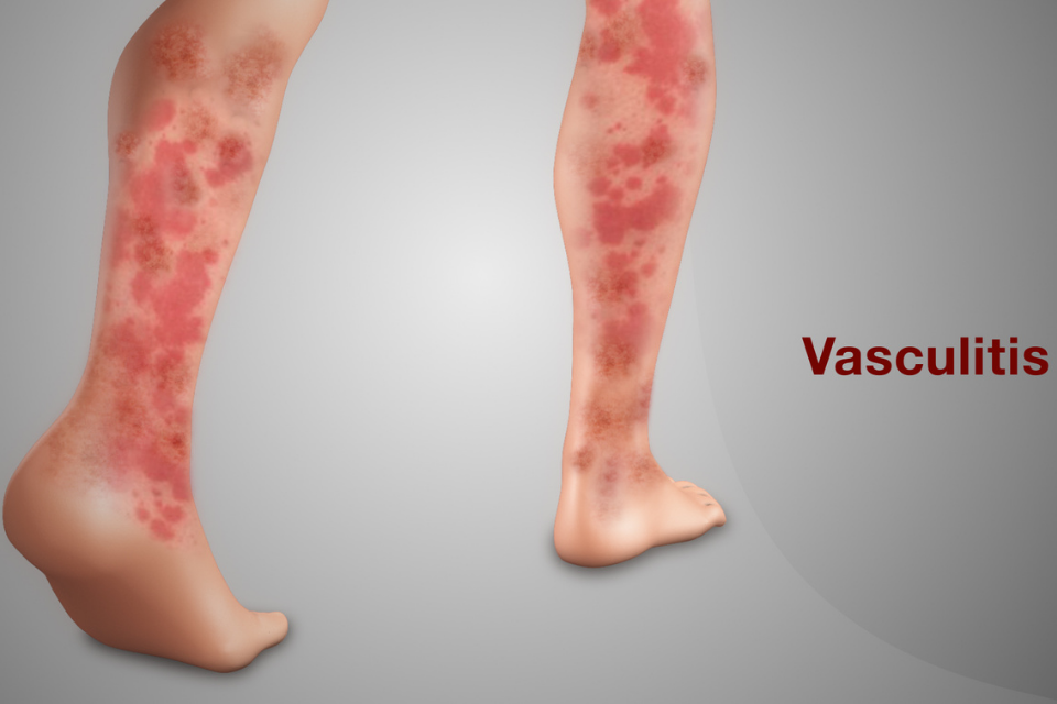
Palpable purpura is pathognomonic for vasculitis. Palpable purpura consists of elevated, nonblanching erythematous lesions. Not all vasculitis presents with palpable purpura, however, and a variety of dermatologic lesions may be seen including macules, papules, wheals, vesicles, bullae, and ulcers. Specific clinical manifestations reflect the size of the involved vessels. Small vessel vasculitis commonly presents with purpura, erythema, urticaria, vesiculobullous lesions, superficial ulcers, and splinter hemorrhages. Medium-vessel vasculitis can present with subcutaneous nodules, erythematous nodules, deep ulcers, livedo reticularis, pitted palmar scars, gangrene of the digits, or infarcts. Review of vital signs may show hypertension if there is renal artery involvement, and thorough neurologic exam may reveal deficits consistent with mononeuritis. Large vessel vasculitis often has no skin manifestations; common findings on physical exam include asymmetric blood pressure, absence of pulses, and bruits.
The important finding of retiform purpura deserves special mention. Retiform purpura consists of nonblanching erythematous lesions in a reticulate, branching, serpentine, or stellate pattern. Retiform purpura is caused by complete loss of blood flow in the dermal and subcutaneous blood vessels. It may be caused by vasculitides such as cryoglobulinemic vasculitis, connective tissue disorder-associated vasculitis, polyarteritis nodosa, microscopic polyangitis, Wegener’s granulomatosis, and Churg-Strauss syndrome. It may also be caused by vasculopathies. The differential diagnosis includes antiphospholipid antibody syndrome, protein C or S deficiencies, disseminated intravascular coagulation, coumadin necrosis, heparin necrosis, cholesterol embolization, calciphylaxis, and type I cryoglobulinemia.
Pathologic evaluation
Biopsy confirms the diagnosis of vasculitis and identifies the size of the involved vessel. In order to obtain the most information, the most purpuric, erythematous, and tender skin should be biopsied. It is also important to biopsy lesions that are less than 48 hours old; after 48 hours lymphocytes and macrophages replace initial inflammatory cells regardless of the underlying type of vasculitis. This is also true for direct immunofluorescence specimens as the likelihood of finding immunoglobulins decreases after 48 to 72 hours. One must also take care when deciding on the type of biopsy to perform. The subcutaneous tissue must be included if a medium-sized vessel vasculitis is suspected. A deep biopsy of the central white area should be performed for livedo reticularis. In cases in which ulcers are present, one should attempt to biopsy nonulcerated sites or the edge of a superficial ulcer. In cases in which deep ulcers are present, biopsy of the subcutaneous tissue can be taken from the central portion of the ulcer. In addition to hematoxylin and eosin staining, a sample may be sent for direct immunofluorescence. See Table 9,4 for histologic findings in specific types of vasculitis.
Laboratory and radiologic evaluation
Thorough laboratory evaluation is required in patients with systemic disease or chronic vasculitis. Laboratory evaluation should include complete blood count with differential, blood urea nitrogen and creatinine, liver function tests, hepatitis B and C serologies, ANCA, ANA, rheumatoid factor, immunoglobulin levels, complement levels, antiphospholipid antibodies, cryoglobulins, urinalysis, and stool for occult blood. In patients with fever and/or a heart murmur, blood cultures and echocardiography may be warranted. Anti-streptolysin O titers can also be checked and may be particularly useful in children. The physician must be aware of the possibility of false negative test results, particularly with cryoglobulins and complement levels. It is suggested that these labs be sent on at least 3 separate occasions.
Treatment
Many cases of cutaneous vasculitis will be self-limited and short-lived. The first step in treatment is to reverse the underlying cause of the vasculitis, if possible. Symptomatic treatment with antihistamines, aspirin, nonsteroidal anti-inflammatory drugs (NSAIDS), leg elevation, ice packs, and avoidance of tight-fitting clothing may be sufficient. In a study of 95 patients with hypersensitivity vasculitis, 54 patients did not require treatment, and another 26 cases resolved with NSAIDS only. It is important to note that NSAIDS have limited value in vasculitides with renal involvement. These agents cause vasoconstriction of the afferent arteriole which decreases renal perfusion and can exacerbate renal failure. In patients with itching or burning or in those with refractory or recurrent skin disease, dapsone (titrated from 25 to 50 mg daily), colchicine (0.5 to 0.6 mg twice daily or three times daily), or pentoxifylline (400 mg three times daily) may be tried.
Systemic immunosuppression may be required in patients who develop systemic symptoms, extensive disease, or persistent lesions. Agents that may be used include prednisone (15 to 80 mg daily or 1 to 1.5 mg/kg daily), methotrexate (5 to 20 mg weekly), azathioprine (50 to 200 mg daily or 0.5 to 2.5 mg/kg daily based on thiopurine methyltransferase (TPMT) level), hydroxychloroquine (400 mg three times daily), mycophenolate mofetil (2 g daily), cyclosporine (2.5 to 5 mg/kg daily, divided twice a day), or cyclophosphamide (2 mg/kg daily) (15, 71).
Therapy for the ANCA-associated vasculitides has been studied in depth. Before the use of cyclophosphamide, Wegener’s granulomatosis was a fatal disease with death typically occurring within 5 to 12 months of disease onset. The introduction of cyclophosphamide and corticosteroids increased survival to 80% (72). A randomized controlled trial of 149 patients with ANCA-associated vasculitis showed that pulse cyclophosphamide (15 mg/kg every 2-3 weeks) was as effective as daily oral cyclophosphamide (2 mg/kg) in inducing remission; in addition, fewer cases of leukopenia were seen with the pulse regimen.
In some patients, methotrexate may be an alternative to cyclophosphamide for induction of remission. A randomized trial of 100 patients found similar remission rates at 6 months of therapy (methotrexate 89.8% and cyclophosphamide 93.5%). However, remission was delayed in patients with lower respiratory tract involvement or more extensive disease. Patients treated with methotrexate had a higher rate of relapse (69.5%) than patients treated with cyclophosphamide (46.5%), and relapse occurred sooner in those treated with methotrexate (13 months) than in those treated with cyclophosphamide (15 months). In this study both treatments were tapered and stopped by 12 months; the high rates of relapse lead the authors to conclude that therapy should be continued longer than 12 months (74).
Cyclophosphamide may be associated with serious adverse effects such as infection, bone marrow toxicity, cystitis, transitional cell carcinoma, myelodysplasia, and infertility (72). An important study by Jayne et al showed that exposure to cyclophosphamide could be safely reduced by substitution of azathioprine after remission was achieved. In this study, comparable rates of relapse were seen in patients who continued on cyclophosphamide after remission (13.7%) and those who were switched to azathioprine (15.5%). Methotrexate has been shown to be an alternative for azathioprine in maintenance therapy. A study by Pagnoux et al showed that the two drugs had similar rates of adverse events (azathioprine 29/63 patients, methotrexate 35/63) and relapse (azathioprine 23/63, methotrexate 21/63). Of note, 73% of these relapses occurred after the study drugs were discontinued. Mycophenolate mofetil appears to be less effective than azathioprine for maintenance of remission. In a study of 156 patients, relapse was seen in 30/80 patients treated with azathioprine and 42/76 patients treated with mycophenolate mofetil (p = 0.03).
Novel biologic therapies targeted against specific components of the immune system may be used in patients in whom conventional therapy has failed. Agents such as infliximab (chimeric anti-tumor necrosis factor-alpha (TNF-alpha) monoclonal antibody), etanercept (fusion protein of the p75 TNF-alpha receptor and IgG1), adalimumab (fully humanized IgG1 anti-TNF-alpha monoclonal antibody), rituximab (chimeric anti-CD20 monoclonal antibody), anakinra (recombinant interleukin-1 receptor antagonist), and IVIG may be used in refractory disease. Prospective studies of infliximab and adalimumab combined with standard therapy of cyclophosphamide and corticosteroids in patients with ANCA-associated vasculitis have shown that these agents may reduce corticosteroid requirements. Mean prednisolone doses decreased from 23.8 mg/day to 8.8 mg/day at week 14 with infliximab. In patients with ANCA-associated vasculitis and renal involvement, adalimumab reduced mean prednisolone doses from 37.1 mg/day to 8.1 mg/day at week 14. A randomized controlled trial of 174 patients found that etanercept was not more effective than placebo in maintenance treatment of Wegener’s granulomatosis. Sustained remission was seen in 69.7% of patients treated with etanercept and in 75.3% of patients in the control group (p = 0.39). A recent study by the European Vasculitis Study Group found that rituximab and cyclophosphamide-based induction regimens for ANCA-associated renal vasculitides had similar rates of sustained remissions and adverse effects. Sustained remission was seen in 76% of patients in the rituximab group (treated with glucocorticoids, rituximab for 4 weeks, and 2 cyclophosphamide pulses) and in 82% of patients in the control group (treated with glucocorticoids and cyclophosphamide for 3-6 months followed by azathioprine). Severe adverse effects occurred in 42% of patients in the rituximab group and 36% of patients in the control group; 18% of patients in each group died. The RAVE-ITN Research Group found that rituximab may be superior to cyclophosphamide for inducing remission in relapsing disease. Disease remission at 6 months (without corticosteroids) was seen in 67% of the rituximab group versus 42% of the control group treated with cyclophosphamide (p = 0.01).
Conclusion
The vasculitides are a heterogeneous group of diseases. While some forms of vasculitis are limited to the skin, the physician must always consider the possibility of systemic involvement. A thorough history, physical examination, and biopsy increase the likelihood of making a correct diagnosis. While some forms of vasculitis progress slowly, others can lead to fatal complications rapidly; the prompt recognition of vasculitis and determination of extent of disease are crucial so that appropriate therapy can be initiated.