Dysplastic nevi are a topic of great debate among dermatologists and one of the staple conditions we treat here at our offices. Normal moles are easy to call and melanoma is easy to spot, but what about in between these conditions? Skin cancer is not a light switch, on/off, but rather a spectrum of atypia leading up to melanoma. This “in-between” phase is a grey area that’s not entirely normal, but also not irregular enough to be called a melanoma. This grey area is known as dysplasia or atypical moles:
The diagnosis of a dysplastic mole is made by a dermatopathologist reading a biopsy of a mole that didn’t look quite right to us. What’s that mean? Well, most of our patients will identify a suspicious mole well before we do. That’s because moles that turn atypical (called “dysplastic” moles) do so very prominently. Unlike some emerging cancers in your body that can grow undetected for years, skin cancer is usually immediately recognizable. Atypical moles do not “hide” well at all and you are the first line of defense to catch an emerging skin cancer before it gets serious and even potentially life-threatening. Because the skin is so immediately accessible and no x-rays or MRIs are needed to see it, its infinitely easier to catch a skin cancer before is gets out of hand.
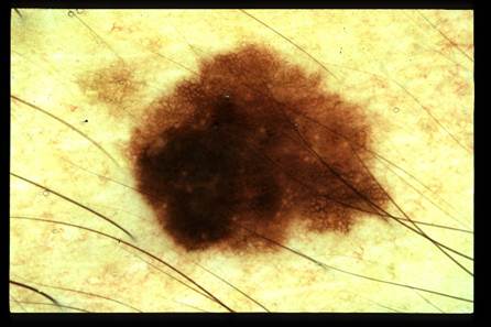
Well, the classic teaching for identifying skin cancer moles is based off of the acronym. A.B.C.D. We’ll begin our discussion there, but there are a couple additional points you’ll want to remember to make sure you’re an at-home expert.
A-Asymmetry. The first thing to look for is a mole that looks different on one side verses another.
B-Border: Look out for moles that have jagged or irregular edges require further examination.
Notice if the mole has very indistinct margins. It would be difficult to trace them out with a ball-point pen. C-Color: moles that have 3 or more colors are begging for attention.
If the mole has nearly 4 colors it is highly atypical. D-Diameter: moles that are larger than 6mm classically should be watched closely. Let’s amend this one to expanding Diameter, that is, if you notice a mole is growing rapidly it should be tested. But if it’s been 10mm since you were in gradeschool, the likelihood of this mole being atypical is very unlikely.
More generally, dermatologist’s follow the “ugly duckling” rule. Finding moles on your body that look different from the others are the same moles we as dermatologists will notice. Let’s say all of your moles are 2-toned with borders that are a bit smudged, but one has 2 colors and perfectly symmetric. Even though its symmetry is reassuring, it’s different from all the other moles your body has made. That’s the mole that is more likely to be atypical under the microscope. Finally, any mole that is symptomatic, whether itching or painful or tingly or burning – any symptom at all, is one that requires a skin biopsy from your dermatologist.
Atypical moles are believed to be pre-cancerous, but just how pre-cancerous we’re not entirely sure. Some of these atypical moles may become cancerous in 500 years (long after you’re gone), but others may do so over several years. Currently, the standard of care is to sample these moles to assess just how atypical the cells are. A skin biopsy can be performed with any board-certified dermatologists and takes less than 5 minutes. It’s almost painless because of a local anesthetic that’s used to numb the area and leaves a tiny scar. If a mole is examined under the microscope and found to contain rapidly-dividing cells or cells that contain irregular DNA patterns, then a complete removal is recommended. If the microscopic atypia is unimpressive then no additional removal is required.
Exciting new technology will soon allow dermatologists to look at the cells that make up your mole in real time without a biopsy. A process known as confocal microscopy allows your doctor to place a hand-held computer flush against your skin to determine a mole’s behavior without a biopsy. The technology is not quite perfected yet, so for now we still need to take a small piece of skin in order to give you an accurate diagnosis regarding that pesky looking, asymmetric, tri-colored, irregularly bordered, itch mole you’ve been trying to ignore.